Management of peroneal nerve involvement after ankle sprain
 IMTA Manager
Happy New Year to all IMTA Blogger
Maitland and Taping (3)
OOOPS sprained your ankle? At yesterday’s new year’s eve city run or gala dinner and dancing? While walking with some %o through the night? Well, you are not the only one….
Background
Twisted or sprained ankles are the most common locomotor system trauma (Garrick 1977, Collins & Comstock 2008, Davidson et al. 2008 in Langendoen et al. 2106).
Recurrences are common and an increasing laxity, up to real instability, may occur. Therapeutically it is the main aim to prevent such a development (Hertel 2008, Santos & Liu 2008, Van Rijn et al. 2008 in Langendoen et al. 2106).
There can be lack of stiffness of the periarticular structures, or a positional fault of the Fibula (or Talus or Os cuboideum) may explain persistent complaints (passive subsystem of stability) (Panjabi 1992 in Langendoen et al. 2106).
Relevant muscles, such as Mm. peronei and or M. tibialis posterior, may be insufficient (active subsystem) (Gutierrez et al. 2007, Hertel 2008 in Langendoen et al. 2106).
The control subsystem may demonstrate dysfunctions like injury related (directly or indirectly by the fibular mechanical interface) neurogenic pain and / or a reduced conduction speed as a logical consequence of immobilization, which offers a rationale to start active rehab as soon as possible.
Testing for neurogenic pain component
Compare Inversion INV and standard SLR left and right, then compare SLR with INV prepositioning. Only the latter usually reproduces the typical pain and shows a significant left-right difference.
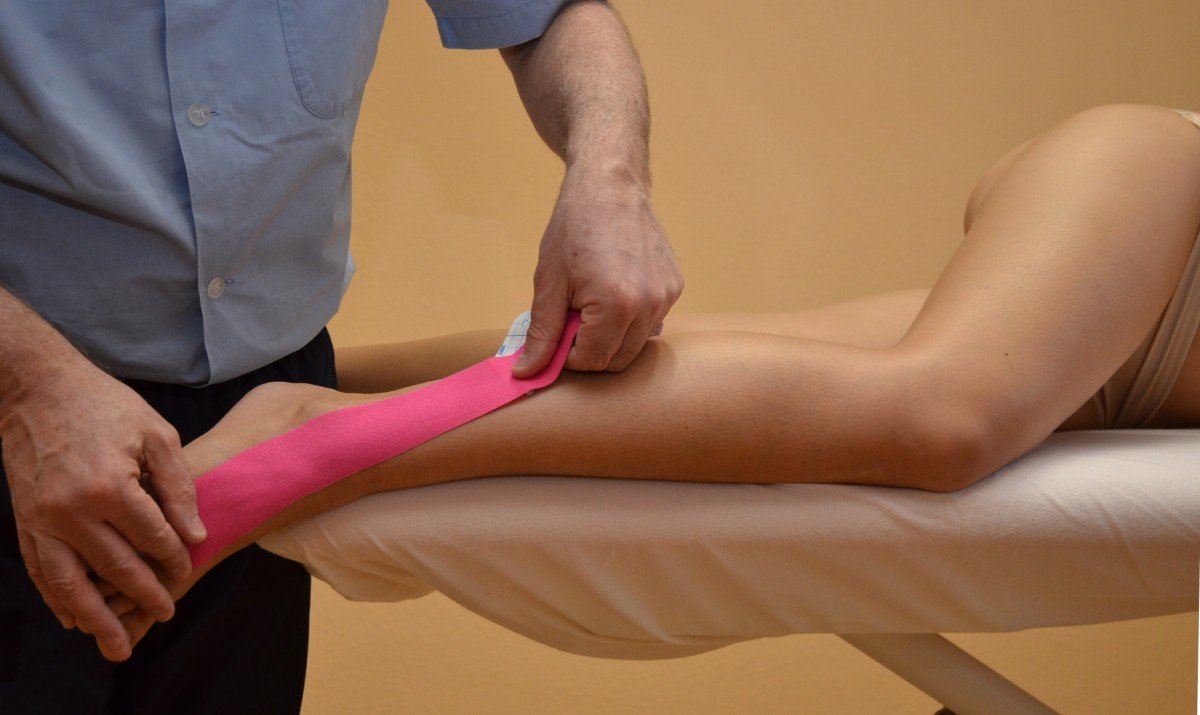
Taping as a conjunct to peroneal nerve slider training
Apply one tape in analogy to a distal pain free peroneal nerve slider exercise: Knee Extension (with Foot DF) alternating with Foot Inv (with Knee F) with the Hip in F/ad in SL position
Starting position 1: side lying with the lower leg at the edge of the plinth, the knee in flexion and the foot in inversion over the edge, all short of onset of pain (P1), but no farther than 1st resistance (R1)
Base 1: anterolateral foot at metatarsal …
Starting position 2: side lying with knee extension short of P1, but no farther than R1 or 30° of flexion, and the foot in eversion
Base 2: distal posterior thigh in the midline, crossing the knee diagonally to the posterior side of the fibular head
Course: remove the paper in knee flexion and foot eversion, apply middle part of the tape during knee extension and foot inversion, check & correct the app and reassess INV+SLR.
Then instruct the patient to do the slider exercise with the tape app till the next session.
(Langendoen et al. 2103, Duran Araneda & Sánchez Maulén 2014)
Good Luck, Feluz ano 2017 & Cheers!,
John & Liz
References
Langendoen J, Sazegar E & Timpe T 2016, Therapeutic Taping Course Manual Lower Quarter, Kinetic Int Hamburg
Langendoen J, Sazegar E & Timpe T 2013, Neurodynamic Taping, Kinetic Int Hamburg
Duran Araneda E, Sánchez Maulén RC 2014 Impaired neurodynamics and the effect of fibular nerve taping application in adult female football players of Colo-Colo, Palestino and Audax Italia. Bachelor Thesis. Universidad Bernardo O’Higgins, Santiago de Chile


Comments